I'm involved in everything from a nutraceutical company to a pharmaceutical company to a medical device company. My whole world revolves around health, and I feel it's my responsibility, in a way, and I say it this way, and I don't take this lightly. - Montel Williams.


Mohammed Arafat | 3rd February, 2022

A new research conducted by Medical University of Vienna stated that a new MRI technique could improve diagnosis and treatment of multiple sclerosis.

To  delay the progression of the disease, it is important that multiple sclerosis (MS) is diagnosed and treated early. Magnetic resonance imaging (MRI) plays an important role in this process. A new MRI method was developed in a university in Vienna as part of a research project that could open a way to quickly diagnose disease function in MS. The study was conducted by a team of researchers led by Wolfgang Bogner at the Vienna university Department of Biomedical Imaging and Image-guided Therapy.

Multiple sclerosis is a systemic disease that appears to change ulcers, especially in the brain. Like many other diseases, there is no cure for this disease, but it can be successfully treated. Early diagnosis is essential for prediction, with highly detailed imaging techniques playing a major role. Although routine MRI can detect brain lesions, scientists are studying ways to detect changes in the microscopic or biological classification of living organisms. 

A method known as proton MR spectroscopy has been identified as a promising tool for this purpose. Using this process, a research team led by Eva Niess and Wolfgang Bognerand other scientists from the Department of Neurology used 7-tesla magnetic MR spectroscopy to compare neurochemical changes in the brain of 65 MS patients with those of 20 healthy controls.

Using 7-tesla MRI, MedUni Vienna researchers have now been able to identify MS-relevant neurochemicals, i.e. chemicals involved in the function of the nervous system. "This allowed us to visualize brain changes in regions that appear normal on conventional MRI scans," says study leader Wolfgang Bogner, pointing to one of the study's main findings. According to the study's lead author, Eva Niess, these findings could play a significant role in the care of MS patients in the future: "Some neurochemical changes that we've been able to visualize with the new technique occur early in the course of the disease and might not only correlate with disability but also predict further disease progression."

Using 7-tesla MRI, the researchers are now able to identify MS-related neurochemicals, i.e. chemicals involved in the functioning of the nervous system. ""This allowed us to visualize brain changes in regions that appear normal on conventional MRI scans," said study leader Wolfgang Bogner, pointing to some of the key findings of the study. 

Niess and Bogner explained that further research is needed before these findings can be incorporated into clinical plans. They say the results showed that 7-tesla spectroscopic MR imaging is a new and important tool in diagnosing multiple sclerosis and treating MS patients.

Looking to the future, Wolfgang Bogner said that If the results are confirmed in further studies, this new neuroimaging technique could become a standard imaging tool for initial diagnosis and for monitoring disease activity and treatment in MS patients.

It’s worth mentioning that the method is currently only available on the 7-Tesla MRI scanner in Austria at MedUni Vienna and only for research purposes. However, the scientific team is working on refining the new method for use in routine clinical MRI scanners.


Mohammed Arafat | 27th January, 2022

Arab Health - Dubia,

After three years of research and almost £15 million (Dh74m) of investment, a Northern Ireland medical company stated that a headset that delivers small electrical pulses to the brain and is worn for just one hour a day could be an inexpensive alternative to treating diabetes and obesity.

The company has said the device could soon offer a non-invasive alternative to medication or brain implant surgery, and that it could be used to target diabetes and metabolism through harmless electric pulses.

Neuromodulation is a science that stimulates the senses to produce a natural biological response, which can be done by using small doses of stimulation directly somewhere in the brain.

The Vestal DM device is said to improve the part of the brain that protects against type 2 diabetes and obesity by controlling blood sugar and appetite.

Dr McKeown, the neuroscientist who has received a £2 million (Dh9m) UK government innovation grant for his research said that the device stimulates the brain to regulate body control, such as respiration, heart rate and blood sugar.

“If those areas are not working well, the brain requires stimulation that is usually delivered via an implant and an expensive surgery that typically costs around $30,000.” He added.

He also mentioned that it has a low-risk option to treat diabetes, considering around a billion people worldwide have either diabetes or pre-diabetes.

With a cost of $500, the device targets the brain, spinal cord and peripheral nerves via applied electrodes. This happens by sending signals into the hypothalamus region of the brain that regulates the body’s metabolic processes like a thermostat, and then the results can be tracked via a mobile app.

A trial that lasted 15 weeks tested the device on 30 Indian patients with type two diabetes. They used it for about five hours a week over a three-month period, to see how effective it was in reducing blood sugar levels.

Now that the device is tested and displayed, the company is seeking US Food and Drug Administration approval. 

Mohammed Arafat | 21st January, 2022

It happened to all of us, and we hated it. This month, COVID-19 became a “party pooper” again as it caused anin-person medical imaging conference to be delayed. The Association for Medical Imaging Management (AHRA) shelving its planned show in Savannah, Georgia, next month.


AHRA Climb was supposed to launch from February 15 to 17 at the DeSoto Hotel in the city’s historic district. However, challenges presented by the new variant of the virus changed the plans. hospital staffing shortages, rising patient admissions, limited testing and uncertain travel scenarios have forced the association to make the decision.


AHRA said in a statement that hosting an in-person event in the current climate is not in the best interest of the imaging community. It hopes to announce revised plans soon.


“The decision to change Climb’s in-person format wasn't taken lightly, but we are committed to being responsive to the times, your needs, and above all, your health and well-being, and the same for your staff and patients,” CEO Daniel Kelsey added.


AHRA’s decision comes after multiple other imaging societies also canceled or delayed in-person conferences, after the omicron variant hi communities. Some of the conferences are the ACR-RBMA Practice Leaders Forum, the Society of Nuclear Medicine & Molecular Imaging’s Mid-Winter affair, and the European Society of Radiology’s annual meeting.


Some other conferences were still planned, such as RSNA and Arab Health, a conference Express Systems and Parts Network will attend this year in the United Arab Emirates.

Mohammed Arafat | 17th January, 2022

The wavelength of visible light ranges from 400 to 700 nm in the visible spectrum. X-rays are considered high-energy with a wavelength of less than 0.0110 nm (100,000 eV), invisible to humans. At present, high-intensity radiation methods are much needed, so the development of new methods is important. Some of the traditional methods include X-ray films, phosphor-based machines, semiconductor-based machines, and gas-based machines.

Many researches were conducted in the last decade about the prospect of semiconductor nanowires (NWs) as an integral part of nanoscale devices and electrical circuits. Three different aspects of the NWs have encouraged previous research.

Second, their high local content enhances their contact with nature, making them more sensitive. The elastic deformation relaxation in the external environment also empowers the formation of new heterostructures that are impossible in planar geometry. Finally, their properties are strongly influenced by their shape, which makes them suitable for use as polarization-dependent sensors.

Materials used for X-ray imaging can be prepared as nanoparticles, nanocomposites or transparent nanoceramics. Using these items for x-ray imaging has different effects; some of which are:

Quantum Confinement Effect

More impact

Structure Effect

The Role of Nanowires

The use of synchrotron-based sensors in the cross-energy spectrum has many potential and offers a variety of benefits, including depth of soil information, element and orbital clarity, and rapid detection of K-absorbing edges and X-ray fluorescence exit methods, medium, and simple elements.

As a result, current forms of synchrotron radiation, such as X-ray microdiffraction techniques, have played a major role in the rapid evolution of nanowire technology. In addition, lens methods were used to analyze ZnO NWs using Bragg coherent diffraction imaging.

The ability to track full stress tensors in 3D with nanometric accuracy has the potential to be very useful in studying complex nanostructures. However, there are a few major drawbacks in this case, because the information related to the NW event is limited.

Most synchrotron processes are now done in NW frameworks, with data obtained at a local scale on large scales. 

The use of nanomaterials for X-ray detection is limited compared to other nanomaterial applications. Exciting techniques, scintillator novel materials, and the actual use of chemicals are all involved in X-ray detection research using nanomaterials.

Mohammed Arafat | 12th January, 2022

Regional anaesthesia device to be demonstrated at Arab Health

The UK technology, which aims to transform regional anesthesia into a one-stop operation and improve patient safety, will participate at Arab Health 2022.

The procedure, SAFIRA, was performed at a theater located in the UK Pavilion by Dr. Emad Fawzy, an anesthesiologist at the Sheikh Khalifa Medical City's main hospital in Abu Dhabi. SAFIRA was developed by medical equipment company Medovate in partnership with doctors from the NHS.

The award-winning device gives anesthesiologists full control of the injection, as well as boosting self-confidence by using a built-in safety mechanism to automatically reduce injection pressure below 20psi, thus helping to reduce the risk of nerve injury to patients.

The live exhibition will take part in the UK theater as part of a series of retrospective surgical simulations. Dr. Fawzy, who previously worked at Queen Elizabeth Hospital King's Lynn NHS Foundation Trust in the UK as part of a team of medical professionals behind the first device, will showcase the device and share his experience of introducing new pioneer technology into the medical profession. .

Dr. Fawzy said that this device is already in use by doctors around the world and the response has been overwhelming, including reports of how technology is helping, hydro-dissection and reducing the amount of local anesthetic needed for effective prevention. 

“It is a great honor to be able to show such a new machine in the Middle East.” He added.

Recent research has sparked a new interest in regional anesthesia worldwide with evidence showing improved clinical outcomes. During the COVID-19 epidemic, both European and American Societies of Regional Anesthesia both produced joint COVID-19 recommendations stating that regional anesthesia should be preferred over conventional anesthesia as the need to reduce aerosol production processes (AGPs) we started.

As well as getting information about SAFIRA, visitors to the Medovate stand will also be able to find out more about the company and its unique portfolio of new medical equipment, all made with doctors from the NHS.

Arab Health takes place on 24 January - 27 January 2020 at Dubai World Trade Center, Dubai, UAE. SAFIRA exhibitions, led by Dr Emad Fawzy, will be held on Tuesday 25 January 11-30 - 12-30.


Mohammed Arafat | 6th January, 2022

The global market for computed tomography (CT) scanners and equipment is expected to drop from $ 7.97 billion by 2020 to $ 7.86 billion by 2021 with a combined annual growth rate (CAGR) of 1.4%. This decline is mainly due to the postponement of various treatments due to the COVID-19 outbreak which has led to a decline in demand for equipment. The market is expected to reach $ 9.26 billion by 2025 at the CAGR of 4.2%.


Leading players in computed tomography (CT) scanners and equipment market are GE Healthcare, Koninklijke Philips NV, Hitachi Ltd., Siemens, Canon Medical Systems Corporation, Neusoft Medical Systems, Hitachi Healthcare America, Neurologicala, Samsung Electronics, and Shimadzu Corp . 


Radiation effects and an increase in health concerns affect computed tomography (CT) scans and a growing market for equipment. Imaging techniques use CT scans that release electromagnetic waves or particles called 'ionizing radiation'. These radiation from man-made sources such as CT scans, nuclear drugs and PET scans carry significant health risks and risks. Low doses can cause cancer over time.


Cancer is the leading cause of death worldwide, with an estimated 9.6 million deaths in 2018. When doses exceed certain levels, they cause skin burns and severe exposure to radiation. According to a national study conducted by the National Council on Radiation Protection and Measurements (NCRP), 2390 people were exposed to radiation, and 190 died.


In 2016, a regulation of radiation protection-computed tomography was developed by the Department of Health (DOH), Washington, which has been in effect since July 2017. By regulation, manufacturers of CT scans are not allowed to make protocol or other software changes. any improvements that affect radiation exposure or image quality.


They need the approval of a leading CT interpreter, specialist, or medical professional to allow for appropriate changes. If there is any uncertainty in the ionizing emission device, it should comply with the given performance standards related to mechanical configuration and other important metrics.


Therefore, manufacturers of medical imaging devices should reduce their ionizing radiation levels effectively to reduce costs, and reduce the warranty periods. Strict rules, such as these, may affect computed tomography scanner devices and the equipment market.


The growing risk of cancer and heart disease is driving the market for computed tomography (CT) scanner and equipment market. According to the Centers for Disease Control and Prevention in the USA, the number of young people with cancer will increase by 24% for men and 21% for women by 2020 than in 2010.


The growing demand for early detection and prevention of these life-threatening diseases, increases the need for appropriate diagnostics that puts them as an opportunity for CT scan manufacturers. 


Companies in the industry combine medical imaging with IT healthcare technologies by providing digital access to improve patient safety, improve health care environment and save patient time and money. Data from CT scans is integrated with an image processing system and a communication system, such as an archive and a communication system (PACS) that integrates CT images from multiple systems into a single interface.


This allows radiologists to safely store, digitally transmit electronic images and related reports, and use photographic data to provide accurate diagnoses. These are increasingly compiled and used for clinical analysis, diagnosis and treatment in departments such as radiology, nuclear medicine imaging, cardiology, pathology, oncology and dermatology.

Mohammed Arafat | 22nd December, 2021


A component of Philips Azurion Image Guided Therapy System, Philips SmartCT application software was announced by Philips Healthcare after its 510(k) clearance was issued by U.S. Food and Drug Administration (FDA).


It  provides interventionalists with CT-like 3-D images using cone beam CT from a sweep of the angiography system C-arm to support diagnosis, therapy planning, treatment and follow-up for interventional cardiology or radiology cath lab procedures. 


SmartCT helps to improve clinical confidence, smooth out workflow, and increase productivity. It also brings precise touch screen controls for enhanced 3-D image acquisition, visibility, vessel / organ separation, and quantity measurements on the side of the table, within the barren area of ​​the intervention lab. It also integrates important laboratory systems and tools needed for complex intervention procedures in an uncluttered laboratory environment where interventions can focus on treating the patient rather than being interrupted by technology.


SmartCT fully integrates Azurion platform control over a touch screen tablet located next to the radiology table. This eliminates the need for doctors to leave the field blank and enter the control room nearby, as well as to support decision-making quickly and efficiently.


“A key part of our image-guided therapy strategy is to combine high-quality, low X-ray dose imaging with a superior user experience that allows interventional radiologists to diagnose and treat patients as part of smoother, safer and less interrupted workflows,” said Ronald Tabaksblat, general manager image guided therapy systems at Philips. 


He added that Philips SmartCT is a major step forward in 3-D imaging, enhancing confidence in the interventional suite and supporting key elements of the quadruple aim of better patient outcomes, enhanced patient and staff experiences and lower cost of care.


“Changing to a new technology can be challenging, but if the system itself can show you the way, it makes it much easier to adopt new advances,” Prof. Hicham Kobeiter, chief of radiology and interventional radiology, Hôpitaux Universitaires Henri-Mondor in Créteil, France. 


Prof. Hicham stated that the new software leads through each step of the procedure, bringing us more confidence and more precision across cardiovascular, oncology and emergency cases.


With SmartCT, users are guided through the image acquisition process and can review and collaborate with CT-like 3-D acquired images in a table-side touch screen module using 3D visualization and measurement tools. These tools are designed to support procedures in a wide range of clinical settings, including neurology, oncology, and cardiac procedures, and incorporate two precise two-point measurements into 3D images, the ability to remove structures from images that impede the region of interest. , as well as the ability to select and maintain high projection angles to be remembered during processes.


The Philips SmartCT imaging, imaging and measurement software is an integral part of the next generation of the Philips Azurion Image Guided Therapy System, launched in September 2020, marking an important step forward in improving clinical and operational laboratory performance and enhancing the image role. -directed interventions in the treatment of patients. Azurion has gained rapid worldwide discovery, demonstrating accelerated non-invasive surgical trends due to its benefits in terms of reducing patient trauma, shorter recovery times and hospital stay, and lower health care costs.


Mohammed Arafat | 17th December, 2021


To get clearer X-ray scans for medical imaging and security screening, international researchers recently introduced a new nanocomposite approach. The approach absorbs X-rays and then reproduces the captured energy capture as light with almost perfect efficiency. 

A report by phys.org stated that the new method could contribute to the improvement of high-resolution for the said two purposes and could result in efficiency gains in devices that range from light-emitting diodes or LEDs, as well as X-ray imaging scintillators, all the way to solar cells.

Jian-Xin Wang, one of the researchers, said that high-Performance Scintillators comprise mainly of either ceramic that requires tough and costly preparation conditions or perovskite materials with poor light and air stability and high toxicity.

“Organic scintillator materials have a good processability and stability but low imaging resolution and detection sensitivity because of the low atomic weight. So, there is limited absorption of X-ray of their component atoms.” He added.

Omar Mohammed, another researcher, and his colleagues have improved the X-ray capture of scintillators at his lab. They did so by combining them with the so-called metal-organic framework (MOF), Zr-FCU-BADC-MOF, integrating high atomic weight zirconium within highly ordered structures.

When X-rays captured the MOF layer of the nanocomposite, the excited pairs of negatively charged electrons known as "excitons" and the positively charged holes were produced. These energy carriers transferred from the MOF to the organic TADF chromophore, supported by the extremely short distance between them, and the energy was produced as light.

"Our energy transfer strategy promotes organic X-ray imaging scintillators from an almost-dead research field into one of the most exciting applications for radiology and security screening. It also applies to other light-conversion applications including light-emitting diodes and solar cells." Mohammed said. 

On the other hand, Wang emphasised that the direct harnessing of singlet and triplet excitons of the TADF chromophores contributed greatly to its remarkably enhanced radioluminescence intensity and X-ray sensitivity. Efficient energy transfer, which the ultrashort distance between layers, and the direct use of singlet and triplet excited states of the TADF chromophore were key. The material's detection limit was improved even more, reaching more than 140 times lower than a standard X-ray medical imaging dose. Mohammed’s team is planning to further improve the performance of their large-scale X-ray imaging scintillators before they take it to the market.

greatly to its remarkably enhanced radioluminescence intensity and X-ray sensitivity.

Efficient energy transfer, which the ultrashort distance between layers, and the direct use of singlet and triplet excited states of the TADF chromophore were key.

The material's detection limit was improved even more, reaching more than 140 times lower than a standard X-ray medical imaging dose.

Mohammed’s team is planning to further improve the performance of their large-scale X-ray imaging scintillators before they take it to the market.


Mohammed Arafat | 9th December, 2021


Almost everyone in the medical imaging field knows that digital flat-panel detectors (FPD) are used in direct digital radiography (DDR) for the conversion of X-rays to light (indirect conversion) or charge (direct conversion). This is read out using a thin film transistor (TFT) array, according to RadioPaedia. 


Nowadays, FPDs are the most used in clinical X-ray imaging systems. They have enabled radiographers to examine X-rays more quickly. However, they are not suited to the complex shape and geometry of the human body. They are ill-matched to our bodies.


Digital curved-panel detectors? What the heck is that? Well, an international team of scientists has identified key design parameters for making curved X-ray detectors that could lead to better quality, and guess what?  SAFTER X-RAYS!


Surrey University research team, said that depending on flat panels means there is unavoidable distortion around the edges of images. They prevent an accurate registration of the X-ray dose delivered. The team stated that there have been a lot of efforts to create flexible detecto, but unfortunately, they have been unsuccessful due to the, “brittle characteristics of the rigid inorganic semiconductors used to make them.” 


The study, published in Advanced Science, identifies design rules for a special class of ‘inorganic in organic’ semiconductors. By tuning the molecular weight of the bismuth oxide nanoparticle sensitised organic semiconductors to lengthen the polymer chains, researchers and scientists could reach more robust, curved digital detectors with high sensitivity or digital film.


Prabodhi Nanayakkara, lead author of the study and PhD student at Surrey University, said that the curved detector concept has shown exceptional mechanical robustness and enables bending radii as small as 1.3mm.”


“The use of organic or ‘inorganic in organic’ semiconductors is also far more cost effective than conventional inorganic semiconductors made from silicon or germanium, which require expensive crystal growth methods. Our approach potentially offers a significant commercial advantage.”


Professor Ravi Silva, director of Surrey’s Advanced Technology Institute, said that the technology has huge potential in medical applications and other X-ray uses.


“We’re working with a spinout company, SilverRay, and hope to turn this technology into the X-ray detector of choice for high sensitivity, high resolution, flexible large area detectors. This technology will help create a ‘revolutionary’ high sensitivity X-ray detector that is scalable due to the design and materials adopted.”


Mohammed Arafat | 2nd December, 2021


The Express Systems and Parts family had its own booth at the RSNA annual meeting that took place in Chicago last week. Learned a lot, Express staff met a lot of leading companies and manufacturers from all around the world. They connected and reconnected with those they met remotely during the pandemic. 


In addition to many other inventions and services by known gigantic companies, one of the things that really surprised the Express team was the new Precision Imaging Network! Produced and developed by the software company Nuance Communications, the Network is an AI-powered cloud platform that delivers patient-specific data and insights from diagnostic imaging into existing clinical and administrative workflows across the healthcare ecosystem, which leads to helping relieve overburdened physicians.


Unveiled at RSNA 2021, this tool is driven by its PowerScribe and PowerShare solutions, used by 80% of U.S. radiologists and connecting more than 10,000 facilities. It also would connect these stakeholders, deploy AI to bolster diagnostics, increase doc efficiency and lower costs.


Officer Peter Durlach, Chief Strategy Officer at Nuance stated that the tool is sort of an electronic version of a pre-read on the image. He added that it uses technology to put another set of eyes on the vast majority of imaging in the country Instead of relying on the radiologist who is overworked and overburdened and might not even have access to all of the images for each patient.


As we all know, diagnostic imaging is very important in our lives. A study published by the Journal of the American Medical Association stated that more than 80 percent of all hospital and health system visits included at least one imaging study, which means the Nuance Precision Imaging Network will help clinicians and facilitate earlier detection and treatment of diseases.


Nuance runs on Azure, which is a cloud computing service operated by Microsoft, which recently announced its intent to acquire Nuance for nearly $20 billion.


Mohammed Arafat | 24th November, 2021


News Agencies - RSNA 2021 is less than one week away, and of course medical imaging companies are waiting for surprises from international manufacturers. Just a few days before the start of this important event, Royal Philips, Philips Healthcare parent company, is expected to launch new MRI scanners. 

1.5-tesla and 3-tesla MRI scanners will be launched along with software-based workflow enhancements, which are designed to enable radiology facilities to operate more efficiently.

One of these new scanners is the MR 5300 scanner. It extends to a new scanner, which the BlueSeal helium-free operation concept Philips first launched in 2018.

To improve workflow, efficiency, and image quality, MR 5300 features a number of enhancements powered by artificial intelligence (AI). It is able to simplify and automate many complex clinical and operational tasks, which will hugely help professionals and clinicians during complicated operations and tasks.MR 5300 sports a 55-cm field of view, enabling extended anatomical coverage.

The MR 7700 scanner is the other scanner that Philips is proud of. It is a 3-tesla magnet that's not only designed to be suitable for clinical purposes, but for research purposes as well. The company says that MR 7700 has integrated multinuclear clinical capabilities, which will give researchers flexibility for new investigations. 

The functionality of the scanner includes five additional nuclei, giving researchers insight into more metabolic and functional information across anatomies. This also makes multinuclei imaging possible as part of daily MR clinical workflow. 

MR 7700 will be shown at RSNA in Chicago next week while it is still pending FDA clearance.

In addition to those new scanners, Philips will be exhibiting a new AI-powered data reconstruction platform, called SmartSpeed, which will apply the company's Adaptive-CS-Net AI algorithm directly to the source of MRI signals.

SmartSpeed is designed to support 97% of current clinical protocols.

It’s worth mentioning that the Express Systems and Parts team will be attending RSNA 2021 in Chicago. The booth number will be #8046


Mohammed Arafat | 18th November, 2021


Like every year, RSNA(Radiological Society of North America) allows manufacturers to show the world the latest innovations and inventions. December 2020 was the month when General Electric Healthcare Company introduced a robotic angiography gantry that Addresses Ergonomic Issues. Allia IGS 7 system, is the name of the new innovative robotic system, uses AI-optimized dose and imaging settings and enables easier use of rotational angiography.


The Allia IGS 7 angiography system was unveiled at the 2020 Radiological Society of North America (RSNA) virtual meeting last year. It is a laser-guided system, which enables exact positioning in the room and enables advanced imaging and guidance technologies found on fixed-based C-arm systems.


Allia IGS 7 is neither floor or ceiling mounted. It is operating on a wheeled gantry so it can easily be parked out of the way for easier patient access or conversion to open procedures. 


Clinicians and professionals know that about 50% of procedures are performed at working positions where they have poor access to user interface, functions and display. The healthcare company collaborated with clinicians to redesign the image guided therapy suite. They focused on ergonomics, ease-of-use and workflow efficiency. 


Designed to enhance user experience, improve workflow efficiency and increase adoption of advanced image guidance in daily practice, Allia IGS 7 allows users to access their essential functions to make it their room with just one click on the personalized interfaces.


This magica;l robot will enable an optimized ergonomic setup for the user’s clinical needs, even in complex working positions; thanks to the redesigned C-arm.


Stephan Haulon, professor of vascular surgery at Groupe Hospitalier Paris Saint Joseph, Hôpital Marie Lannelongue said that Allia IGS 7 is the result of a real partnership between GE and a high-volume user like him.“


“There has been a real focus on ergonomy, working space, ease of use. What I truly believe is that this image guided system is an assistant to perform all those endovascular procedures. The overall impression of this new generation was excellent.” He added.


Learn more about Allia IGS 7 and other equipments and systems visiting our website or talking to one of our representatives. Don't forget, we will be participating in RSNA annual meeting at booth #8046


Mohammed Arafat | 11th November, 2021


CT scanners technology has completely changed since the 1970s. Most Imaging companies these days offer this innovative GE LightSpeed 16 slices. 

This magical system allowed medical practitioners and others to have great power and lower dose for their patients.


GE LightSpeed 16 slices is one of the most advanced and latest models in CT scanner technology. It gives the doctors and practitioners the flexibility and power they need to perform their work easily. In addition to being the first CT scanner to deliver 16 slices per rotation, it images 0.63 and 1.25 mm slices, and it also images 1, 2, 4, and 8 slice modes. The following are more features of this system:

  1. The 4th evolution in LightSpeed

  2. GE microVoxel® technology

  3. Image quality in three dimensions

  4. Ideal for Complete & Fast Head Scanning

  5. 70cm Gantry Opening

  6. LightSpeed Gantry W/RF Slip-Ring

  7. ± 30 degrees Gantry Tilt

  8. Helical Tilt

  9. Dual High Color, High Contrast 19in. Flat Panel LCD Monitors

  10. Performix 6.3 mHu Tube

  11. 16 Slice Matrix Detector

  12. Patient 16-Slice

  13. Patient Table with Metalless Cradle

  14. Light Speed PDU

  15. High Performance DAS

  16. 53.2kW Generator, 10 to 440 MA

  17. 80, 100, 120, & 140kV Selections

  18. Sub-Second Scanning (0.5 Second Scan Time)

  19. Auto MA to Optimize X-Ray Usage

  20. SMART Start Gantry Activated Scanning

  21. SMART Helical for Exam Streamlining

  22. SMART Prep Instant Contrast Monitoring

  23. SMART Speed Scan Time Enhancer

  24. SMART Tools to Automate Exam Tasks

  25. Variviewer

  26. 120 sec. Continuous Helical & Up to 2000 Contiguous Rotations

  27. 160cm Max. Scan Volume Based on Table Travel

  28. Pitch of Quad 0.75:1 (3:1) or 1.5:1 (6:1)

  29. Magnetic Optical Disk Image Storage


Why wait? talk to us and ask questions regarding this magical system.

Mohammed Arafat from MIT | 4th November, 2021


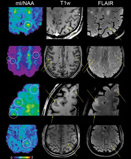
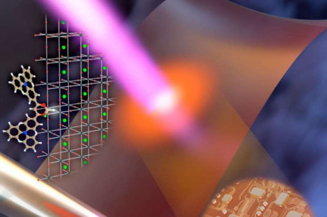
A team at Massachusetts Institute of Technology (MIT) has developed a modified version of two-photon imaging that can scan deeper within tissue and perform the imaging much faster than previously possible.

o create high-resolution, 3D images of tissues such as the brain, researchers often use two-photon microscopy, which involves aiming a high-intensity laser at the specimen to induce fluorescence excitation. However, scanning deep within the brain can be difficult because light scatters off of tissues as it goes deeper, making images blurry.

Two-photon imaging is also time-consuming, as it usually requires scanning individual pixels one at a time. A team of MIT and Harvard University researchers has now developed a modified version of two-photon imaging that can image deeper within tissue and perform the imaging much more quickly than what was previously possible.

This kind of imaging could allow scientists to more rapidly obtain high-resolution images of structures such as blood vessels and individual neurons within the brain, the researchers say.

"By modifying the laser beam coming into the tissue, we showed that we can go deeper and we can do finer imaging than the previous techniques," says Murat Yildirim, an MIT research scientist and one of the authors of the new study.

MIT graduate student Cheng Zheng and former postdoc Jong Kang Park are the lead authors of the paper, which appears today in Science Advances. Dushan N. Wadduwage, a former MIT postdoc who is now a John Harvard Distinguished Science Fellow in Imaging at the Center for Advanced Imaging at Harvard University, is the paper's senior author. Other authors include Josiah Boivin, an MIT postdoc; Yi Xue, a former MIT graduate student; Mriganka Sur, the Newton Professor of Neuroscience at MIT; and Peter So, an MIT professor of mechanical engineering and of biological engineering.

Deep imaging

Two-photon microscopy works by shining an intense beam of near-infrared light onto a single point within a sample, inducing simultaneous absorption of two photons at the focal point, where the intensity is the highest. This long-wavelength, low-energy light can penetrate deeper into tissue without damaging it, allowing for imaging below the surface.

However, two-photon excitation generates images by fluorescence, and the fluorescent signal is in the visible spectral region. When imaging deeper into tissue samples, the fluorescent light scatters more and the image becomes blurry. Imaging many layers of tissue is also very time-consuming. Using wide-field imaging, in which an entire plane of tissue is illuminated at once, can speed up the process, but the resolution of this approach is not as great as that of point-by-point scanning.

The MIT team wanted to develop a method that would allow them to image a large tissue sample all at once, while still maintaining the high resolution of point-by-point scanning. To achieve that, they came up with a way to manipulate the light that they shine onto the sample. They use a form of wide-field microscopy, shining a plane of light onto the tissue, but modify the amplitude of the light so that they can turn each pixel on or off at different times. Some pixels are lit up while nearby pixels remain dark, and this predesigned pattern can be detected in the light scattered by the tissue.

"We can turn each pixel on or off by this kind of modulation," Zheng says. "If we turn off some of the spots, that creates space around each pixel, so now we can know what is happening in each of the individual spots."

After the researchers obtain the raw images, they reconstruct each pixel using a computer algorithm that they created.

"We control the shape of the light and we get the response from the tissue. From these responses, we try to resolve what kind of scattering the tissue has. As we do the reconstructions from our raw images, we can get a lot of information that you cannot see in the raw images," Yildirim says.

Using this technique, the researchers showed that they could image about 200 microns deep into slices of muscle and kidney tissue, and about 300 microns into the brains of mice. That is about twice as deep as was possible without this patterned excitation and computational reconstruction, Yildirim says. The technique can also generate images about 100 to 1,000 times faster than conventional two-photon microscopy.

Brain structure

This type of imaging should allow researchers to more rapidly obtain high-resolution images of neurons in the brain, as well as other structures such as blood vessels. Imaging blood vessels in the brains of mice could be particularly useful for learning more about how blood flow is affected by neurodegenerative diseases such as Alzheimer's, Yildirim says.

"All the studies of blood flow or morphology of the blood vessel structures are based on two-photon or three-photon point scanning systems, so they're slow," he says. "By using this technology, we can really perform high-speed volumetric imaging of blood flow and blood vessel structure in order to understand the changes in blood flow."

The technique could also lend itself to measuring neuronal activity, by adding voltage-sensitive fluorescent dyes or fluorescent calcium probes that light up when neurons are excited. It could also be useful for analyzing other types of tissue, including tumors, where it could be used to help determine the edges of a tumor.


Mohammed Arafat | 28th October, 2021

Medical imaging is an important part in science and medicines, helping provide a high quality of images for the diagnosis of disease. The workstation of such images comprises recording and storing the results of diagnostic with help of software implanted diagnostic devices that help the health care professionals to care for the disease of interest.

According to the report published by the Market Research Store, the global demand for the medical imaging workstations market in 2020 was approximately $4,808.4 Million, but it will grow at a CAGR of 7.2%, reaching around  $7,200.6 Million by 2026.

North American market is estimated for the highest market growth rate in the global medical imaging workstations owing to various factors such as the incidence of an enormous number of imaging centers.

Among the aspects that are propelling the market growth are the launching of technologically higher and inexpensive systems, the developing user preference for digital platforms, the rising public-private investments to support product improvement, the escalating overall burden of target diseases, the establishment of innovative medical facilities, and the augmented product commercialization.

Global Medical Imaging Workstations Market: By Modality

·         Magnetic Resonance Imaging (MRI)

·         Computed Tomography (CT)

·         Ultrasound

·         Mammography

·         Others

Global Medical Imaging Workstations Market: By Component

·         Display Units

·         Display Controller Cards

·         Visualization Software

·         Central Processing Units

Global Medical Imaging Workstations Market: By Usage Mode

·         Thick Client Workstations

·         Thin Client Workstations

Global Medical Imaging Workstations Market: By Application

·         Clinical Review

·         Diagnostic Imaging

·         Advanced Imaging

Global Medical Imaging Workstations Market: By Clinical Specialty

·         Oncology

·         Cardiology

·         Urology

·         Obstetrics and Gynecology

·         General Imaging/Radiology

·         Orthopedics

·         Mammography

·         Other Clinical Specialties

Global Medical Imaging Workstations Market: Regional Segmentation Analysis

·         North America

o    The U.S.

o    Canada

·         Europe

o    France

o    The UK

o    Spain

o    Germany

o    Italy

o    Rest of Europe

·         Asia Pacific

o    China

o    Japan

o    India

o    South Korea

o    Southeast Asia

o    Rest of Asia Pacific

·         Latin America

o    Brazil

o    Mexico

o    Rest of Latin America

·         Middle East & Africa

o    GCC

o    South Africa

o    Rest of Middle East & Africa


Mohammed Arafat | 21st October, 2021


An article published on Research and Market website stated that the medical equipment maintenance market is projected to reach $74.2 billion by the year of 2026 from USD 45.2 billion in 2021. This growth was caused by the growth in the associated equipment market, which is segmented multi-vendor OEMs(Original equipment manufacturers), single-vendor OEMs, ISOs, and in-house maintenance. This market is primarily driven by the increasing number of diagnostic imaging procedures across the globe. A rise in the prevalence of chronic diseases is also expected to propel the use of advanced imaging equipment and subsequently drive the demand for associated maintenance services.


Original equipment manufacturers provide a wide range of maintenance services, such as corrective maintenance, operational maintenance, planned maintenance, etc, with the Preventive maintenance segment accounting for a larger share of the market in 2020.


This type of maintenance is carried out for life support devices/equipment used for diagnosis and treatment, and devices that significantly impact the safety of patients. Examples about that are: defibrillators, ECG machines, pulse oximeters, infusion pumps, and electrosurgical units. Preventive maintenance is critical for equipment present in radiology departments due to mandatory certifications and regulations, and the high cost of equipment. 


The medical equipment maintenance consists of diagnostic imaging equipment, patient monitoring & life support devices, endoscopic devices, surgical equipment, ophthalmology equipment, medical lasers, electrosurgical equipment, radiotherapy devices, dental equipment, laboratory equipment, and durable medical equipment. And this market is divided into CT scanners, MRI systems, X-ray systems, ultrasound systems, angiography systems, nuclear imaging equipment, mammography systems, and fluoroscopy systems.


Mohammed Arafat | 14th October, 2021


During the past two years, the world struggled trying to beat COVID-19 and its aftermath. Thousands died every day, the economy collapsed entirely, local and international flights paused, streets emptied, and offices had no inhabitants. 

Just recently, when the vaccines were made and became available, many regions started to breathe. Life started to come back, but slowly, people started to have smiles on their faces, and spirits started to become more optimistic, thanks to the vaccines companies, first responders, and to the patient people who abided with the COVID regulations set by their local governments.

Although this very important update improved many main sectors in the lives of millions, COVID created shortages of an array of American medical supplies, not only masks, but also other medical parts and equipment needed at medical facilities. 

Many medical experts believe that getting specific types of medical supplies can take up to five months, while it used to take from 3 to 6 weeks before the start of the pandemic.

In addition to local and international shipping being delayed, what helped cause this shortage is the shortages of raw materials, like plastics, metals, glass, and electronics. Many American producers are waiting for parts from overseas, which are jammed in seaports.

A few weeks ago, the Port of Los Angeles/Long Beach stated that a record of more than 60 container vessels were waiting offshore to unload goods, including many that carry raw materials needed for medical products and equipment.

Surprisingly, the shortages in supplies cause higher prices, which fuels fears of a wave of sustained inflation according to an article published by Reuters.

Some of the backlogs, though, are easing, and things are believed to get better over the coming months, but that does not mean the whole issue will be resolved entirely.

Express Systems and Parts Network, Inc, based in Aurora, Ohio, and its staff members are doing their best to be one of the best medical equipment and parts suppliers during these tough times. From a huge warehouse stocked with thousands of parts and complete equipment, to a professional team that knows what they are doing, Express’ goal is customer satisfaction.

Mohammed Arafat | 7th October, 2021

According to a research published on October 2nd by Clinical Imaging, many American students are influenced by concerns over having their potential jobs replaced by artificial Intelligence (AI). Many of those medical students are radiology students across the country.
 
The purpose of the study was to show whether medical students have significant anxiety about pursuing radiology as a career due to fear from AI. The survey was conducted in 32 US medical schools, and 463 students participated in it anonymously. It showed that one out of six students in the US would not choose radiology as their specialty at school because they are scared that AI would replace radiology in the near future.
 
Dr. Kristin Reeder of the Quinnipiac University Frank H. Netter School of Medicine stated that artificial intelligence has a negative impact on American medical students’ choice of radiology as a future career.
 
“This is a phenomenon influenced by both individual concerns and exposure to AI from the medical community.” He added.
 
Surprisingly, when medical students get exposed to AI, either at school or at home, they rank radiology lower. However, when they receive education in AI, they mostly will consider radiology as a career.
 
Reviewing the students’ responses, the researchers found out that half of those students considered radiology as one of their top three choices before the survey was conducted, and less than a third of those students wouldn’t pursue radiology due to fear from AI. This means that the concern toward AI and job decrease was correlated.
 
In addition, the researchers found out that fear from decreased job opportunities and lack of understanding radiology were associated with ranking radiology lower because of Artificial Intelligence.
 
The researchers believe that efforts to include Artificial Intelligence in the medical curriculum in the US are underway in small number.
 
To sum up, although AI has a significant negative impact on US medical students’ choice of radiology as a future career, we all know that it’s clear that artificial intelligence continues to form the future of radiology and what medical students think of it.
 

Mohammed Arafat | 29th September, 2021

Ultrasound imaging is a medical tool that helps a physician/professional evaluate, diagnose, and treat medical conditions, include but not limited to:

·      Abdominal ultrasound

·      Bone sonometric

·      Breast ultrasound.

·      Heart rate monitors.

·      Doppler ultrasound

·      Echocardiogram.

·      Ophthalmic ultrasound.


The functionality of the ultrasound image is very complicated as it’s based on the reflection of the waves of the body structures. In other words, the amplitudes of the sound signal traveling through the body provide the information necessary to produce a medical image.


What are the benefits of ultrasound?

As we mentioned earlier, ultrasound imaging is very essential for daily medical diagnoses. Being the brainchild of engineer Tom Brown and Obstetrician Ian Donald, It has been used for over 20 years, showing an excellent safety record.


What are the risks of ultrasound?

Although ultrasound imaging is considered safe when it’s used by professionally-trained providers, it can cause biological effects on the patient’s body. Its waves can heat the body tissues, produce small pockets of gas in body fluids.

Some American organizations advise prudent use of ultrasound imaging in pregnancy. They suggest less use of it for non-medical purposes. 


How do you prepare for an ultrasound exam?

Some ultrasound exams require some preparation, such as:

·      During some exams, like gallbladder ultrasound, your doctor may ask that you not eat or drink for a certain period before the exam.

·      During Pelvic ultrasound, you may be required to come with a full bladder based on your doctor’s guidance.

·      Children and elders might have special treatments.

An ultrasound exam, which takes from 30 minutes to an hour, is usually painless. However, you may experience mild discomfort as the sonographer guides the transducer over your body, especially if you're required to have a full bladder, or inserts it into your body.

Mohammed Arafat | 24th September, 2021

Since one of its jobs is to de-install complete imaging equipment and systems and units from the fifty states, Express Systems and Parts Network is opening its arms for one of the most popular CTs in the world. SOMATOM Emotion Eco 16-Slice is a customer-focused philosophy Siemens product that is a huge part of the daily clinical routine. 


Whether you are a professional who does business with clinics and hospitals, or a clinician who puts their patient's comfort first, this magical CT's high quality provides uninterrupted patient care and excellent clinical reliability in all of the different circumstances.


While, simultaneously, lowering costs, 16 slice CT is considered one of the most economical CTs, as it has a lower requirement for preventive maintenance.


SOMATOM Emotion Eco 16-slice Configuration Specifications:

Manufacturer: Siemens

System Name: Somatom Emotion 16 (Classic) Square Gantry - RS Eco

Scalable Ref.

Date of Manufacture: 08/22/2003

Serial Number: NA

System Fully Functional? Yes

Condition: 9

Current Service Contract: NA

Console Type: Syngo

Software Version: NA

Software Options List:

Syngo Osteo CT #AWP

Osteo Mattress with Phantom

syngo Dental CT #AWP

WorkStream 4D

Recon Plus

DoseMAP


SOMATOM Emotion Eco 16-slice Configuration Features:

Powerful V-engine gradients (30mT/m)

State-of-the-art zero helium boil-off magnet

Parallel Imaging in all directions and head to toe

Reduce acquisition times with PAT factors up to 8

Water-cooled gradient system with 24mT/m and 55T/m/s slew rate

Increased efficiency with linked database, fast reconstruction, WorkStream 4D, and dose reduction with CARE Dose4D

Less downtime with faster installation.

Reduced scheduled maintenance requirements

More than 98% of the materials used can be returned to the flow of recyclable materials

SafeCT Low-dose Solution

16-slice acquisition

Wide range of clinical applications

Fast and efficient with 0.6 second rotation time

Multislice UFC detector

0.75 or 1.5mm slices per rotation in less than 20 seconds

Small diagnostic detail for acquisition

105 millisecond temporal resolution for clear depiction of blood vessels

Scan any anatomy with a single breathhold

Pathology seen from any perspective

Low patient dose


Interested in learning more? Download the system's manual from 
here.


I agrees and consent to the Terms of Service and Privacy Policy.