I'm involved in everything from a nutraceutical company to a pharmaceutical company to a medical device company. My whole world revolves around health, and I feel it's my responsibility, in a way, and I say it this way, and I don't take this lightly. - Montel Williams.


Express Systems | 24th June, 2022

Medical News Agencies - Researchers are exploring ways to use artificial intelligence in medical imaging. The need for artificial intelligence is constantly growing in the medical imaging software market. From heart events, neurological conditions, fractures, or thoracic problems, artificial intelligence helps physicians diagnose and provide immediate treatment. The use of AI in medical imaging has improved medical evaluation, improved pharmaceutical software, reduced physician burden, etc.

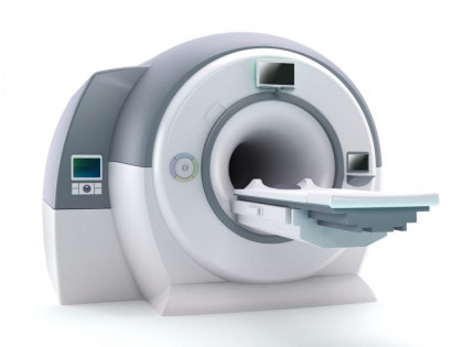
There have been many technological advances in AI-based medical imaging technologies, which have shown their growing popularity in high-income countries. Other improvements include the development of integrated rtiI software, which can be integrated directly into digital imaging (MRI or CT scanner) that facilitates automated medical imaging analysis. Other developments include the integration of smartphone technology into AI into a medical model where advanced health professionals can assess a variety of conditions using a smartphone.


AI in medical imagination has attracted the attention of several radiologists around the world. It provides faster and more accurate results and reduces diagnostic errors at a reduced cost compared to traditional medical imaging methods. Therefore, radiologists believe that AI in medical imaging may present a growing possibility for its increasing implementation in the coming years.


In recent years, many large established companies, such as GE Healthcare and Siemens Healthineers, have been able to grow AI in the medical imaging market by making huge investments to increase partnerships and acquisitions. Other large healthcare or software companies that have not previously invested in health care, such as Thermo Fisher Scientific and Paraxel, have begun investing heavily in the market.

Siemens Healthineers, General Electric (GE) Company, Koninklijke Philips, and IBM Watson Health are major players in the global AI market of medical thought. International players are focused on developing new products with advanced technology and expanding their product portfolio to stay competitive. They continue to invest heavily in R&D to expand their product portfolio. Manufacturers like GE Healthcare are always focused on introducing new products through new technology platforms that open the platform (Edison Developer Program) of other companies that provide artificial intelligence technology to measure and deliver their advanced programs across the GE Healthcare customer base.


Many big players are busy with strategic purchases and partnerships that continue to be a competitive strategy for key players, thus helping them to grow in tandem. The authorization of new products associated with R&D activities helps retailers increase their presence, promote growth, and strengthen their position in the global market. By 2021, more than 30 countries have approved AI for FDA and CE-approved medical imaging technologies.

There is growing support and investment by public and private organizations, including large corporations, which is also one of the major driving forces for AI in the medical thought market. For example, more than 20 newcomers from different regions have received funding to develop AI-based medical imaging technologies.

Hospitals buy software-based intelligence suits as a complete package for use or adopt a single system at a time that is widely used in the industry. The center of imaging imaging is an important source of revenue generation through thought processes, and they are primarily involved in the use of advanced products, which will attract customers. For example, AI in medical imaging, along with clinical data, helps physicians to accurately predict heart attacks in patients.

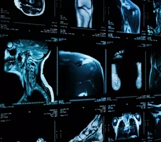
Neurology plays a dominant role in the industry. Much of the product development of artificial intelligence is focused on river processing. These Downstream analyzes mainly include the artificial intelligence of differentiation, anatomical properties, and the measurement of disease scores. Conditions such as intracranial hemorrhage, ischemic stroke, primary brain tumors, cerebral metastases, and abnormal white matter signal, which were industry needs, have become commercially available solutions within the radiology industry.

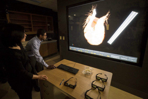
AI in medical imaging, especially cardiovascular magnetic resonance (CMR), is transformed by providing in-depth learning solutions, especially imaging, reconstruction, and analysis, which help support clinical decisions. CMR is an established tool for standard clinical decision making, which includes diagnostic, follow-up, real-time procedures, and pre-procedure planning.

In-depth study methods have provided the greatest success in analyzing medical images. Help them with high accuracy, efficiency, stability, and balance. Artificial intelligence has become a useful medical tool with benefits such as error reduction, accuracy, faster computer use, and better diagnostics. Indigenous language analysis, Computer Vision, and Context-Aware Computing technology are also used to create new analytical methods for medical imaging products.

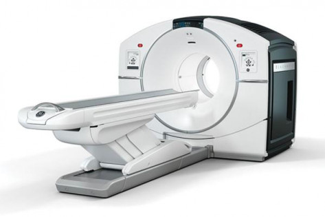
In 2021, Philips unveiled its new AI-enabled CT imaging portfolio. Their new CT 5100 with smart workflow system uses artificial intelligence in every step of processing CT images.

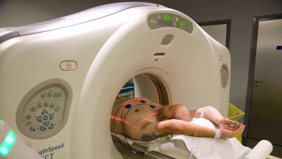
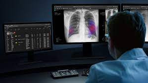
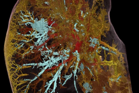
Siemens Healthineers's AI-Rad chest CT detects and highlights lung tumors. Plant load is calculated automatically.
In addition, AI-Rad chest X-Ray played a major role in patient control during COVID-19 violence. The artificial intelligence-Rad file automatically scans X-ray images of straight chest, pneumothorax, nodule

Express Systems | 15th June, 2022

Artificial intelligence (AI) has great potential to improve medical thinking, and AI methods often show more comparable or superior performance than reviews by medical professionals. However, the presentation at the Society of Nuclear Medicine and Molecular Imaging (SNMMI) Annual Meeting highlighted significant risks to AI models, which engineers and users should consider. In particular, presenters highlighted data threats and data fraud.

The authors conducted a review of threats and mitigation strategies to highlight the importance of data security in AI clinical thinking efforts. Among the concerns are productive adversarial networks (GANs): "unregulated neural networks, competing to produce new models in a given training sample."

In the case of photography, the photographs produced are inseparable from the original model. GANs used to create in-depth fake photos and videos. Whether intentionally or not, the process can be manipulated.

Such deception can have far-reaching consequences, with proponents claiming, such as forged images, delays in obtaining problematic images because they appear to be genuine, improper treatment effects if the decision is based on altered images, and financial implications.

They provided a few suggestions for reducing these risks, including image authentication in addition to AI verification, verified image detection watermarks, and encryption across all categories (as images move between scans, storage, workspaces, and more). They also encourage AI developers to educate AI users about issues of trust and trust.

Researchers beleive that effective data security strategies are needed to prevent data compromise while promoting the development of medical imaging technology and patient care through AI methods. It is important that health care professionals are informed of the risks in order to balance patient care obligations with the obligation to control data integrity.

Express Systems | 9th June, 2022

Brainy Insights - The increase in the number of older people and the growing demand for thinking technology in the health care sector is driving the growth of the C-arms market. Lifestyle changes also affect the health status of people leading to chronic diseases such as cancer, respiratory diseases, and others that require surgery for treatment that furthers market growth during the forecast period. However, the high cost of C-Arms equipment due to increased efficiency and improved technology hinders market growth. People these days are increasing their interest in minimally invasive surgery as there are smaller stitches in the body. But smaller stitches limit access to internal organs, which can be accessed using the C-arms. The growing demand for less aggressive surgical procedures is expected to stimulate market growth during forecasting.

To improve their market position in the global C-Arms market, key players are now focused on adopting strategies such as brand design, integration and acquisition, recent developments, joint ventures, partnerships, and collaborations.

C-Arms is a roaming tool used during surgery to scan body parts. The C-Arms device is used to verify the placement of implants. C-arms are portable devices that provide excellent performance and flexibility in surgical procedures. The new C-arms equipment with three-dimensional tomography is a creative process that helps to obtain a more accurate and accurate image of the internal organs. The c-arms device uses a two-dimensional X-ray X-ray device designed to create images similar to CT. The C-arms device sweeps through the patient's body, capturing a few 2D images to aid in surgery.

Essential Findings

● In 2021, the C-arms segment dominated the market with a market share of about 63.4% and a market capitalization of 1273.45 million.

Part of the product type is divided into fixed and mobile. By 2021, the C-arms segment dominated the market with a market share of about 63.4% and a market capitalization of 1273.45 million. C-arms mobile devices are flexible and provide excellent control, which helps surgeons with imaging during surgery.

● By 2021, part of the orthopedics and trauma occupied a large share of the market, with 24% and a market revenue of 482.0 million.

Part of the app type includes bone and trauma, oncology, cardiology, gastroenterology, neurology, and more. By 2021, the orthopedics and trauma segment had the largest market share, with 24% and a market capitalization of $ 482.0 million. Most orthopedic surgery is less invasive and requires C-arms, which promotes market growth in the segment.

● In 2021, half of the hospitals had the largest market share, at 35.6% and a market revenue of 715.0 million.

The proportion of end users is divided into hospitals, clinics, diagnostic centers, and more. By 2021, half the hospitals dominated the market with a market share of about 35.6% and a market revenue of 715.0 million. Hospitals have improved infrastructure to perform all operations and have state-of-the-art medical equipment. As a result, more and more patients are visiting hospitals, which is encouraging market growth.

Express Systems | 3rd June, 2022

An ACR-led initiative recently addressed the issue of incompetence radiologist and recommended follow-up care after accidentally diagnosed diagnosis. The proposed measures were recently published in the Journal of the American College of Radiology.

"Care gaps occur when radiology follow-up recommendations are poorly addressed or incomplete, leading to misdiagnosis or delays that can lead to serious patient outcomes," co-author Nadja Kadom, MD, Director, Radiology Quality, Department of Medicine, and Child Care. of Atlanta, and co-authors wrote.

To address this issue a specialist panel of experts (TEP) was formed to advise quality measures that would improve communication between providers and patients and thus increase the standards for completing follow-up care. The measures are designed to serve as a guide for the process of developing their own communication systems and ways to improve post-accident follow-up that can be acted upon by radiologists.

The foundation for the ACR-led program is based on nine steps — four on outcomes and five on processes.

Suggested outcome measures include the need to close the communication loop upon completion of possible risk assessment recommendations (AIFs), with specific guidance regarding the diagnosis of abdominal aortic aneurysm and pulmonary tumors, in addition to imaging follow-up and monitoring recommendations in cases of acquisition. For each of these steps, a successful follow-up was defined as the completion of the recommended image within 30 days before and 60 days after the recommended follow-up period.

"This initiative serves the main purpose of the initiative, to assess the percentage of patients with at least one AIF who received follow-up images during the recommended period," the authors explained.

The procedural steps included closed communication procedures between patients, radiologists and referral providers in charge of ongoing patient care. A tracking system that incorporates reminders and deadlines was also included in the process estimates.

"This center- and rating scale assesses the percentage of final reports containing AIFs and recommendations for image tracking when the findings are passed on to the patient."

The authors noted the timely arrival of the proposed measures, referring to the CMS prioritization of patient outcomes. Additionally, the measures, which can be applied to a variety of practices as needed, can help close patient safety gaps and provide more insight into proper and timely management of autoimmune findings, experts suggest.

Express Systems | 26th May, 2022

PhysicsWorld - A new type of low-resolution MRI scanner, that does not require dedicated protection, can effectively diagnose stroke and detect blood clots in the brain as small as 4 mm in size. In a study of 50 patients with ischemic stroke treated at Yale New Haven Hospital, intracranial imaging with pMRI detected ischemic infarcts in 90% of patients. The prospective study is the first to show that a portable MRI system of 0.064 T Swoop can be used to directly diagnose and diagnose a stroke near a patient's bedside.

The ability to quickly differentiate ischemic stroke, where inhibition interrupts blood supply to the brain, from the hemorrhagic side, and where there is bleeding in the brain, is essential to accelerate effective treatment. Ischemic stroke, the most common type of stroke, is usually treated with thrombolytic "clot busting" treatment. This method, however, is not suitable for hemorrhagic stroke.

The European Society for Cardiology and the American Heart Association recommend that all patients with a stroke receive a brain scan as soon as they arrive at the hospital to avoid bleeding inside the cranial. CT is the preferred imaging modality to diagnose stroke, in which radiation MRI is becoming increasingly popular; but access to stationary MRI scans can be limited.

Low-level PMRI scans can prove an effective point-of-care diagnosis. The Swoop pMRI scanner, which includes an eight-channel radiofrequency head coil, operates from a common electrical outlet, requires no cryogenics and incorporates electromagnetic interference, eliminating the need for a secure chamber.

Its compact size (140 cm high and 86 cm wide) allows for use in inpatients or emergency department settings, and does not require specialized MRI specialists to operate. Importantly, pMRI is unaffected and does not endanger the operation of nearby hospital equipment.

A portable, bedside MRI scanner solution opens the doors to rethinking how to deliver quality care, reachs patients and communities around the world, and better understands the basics of emotional and physical damage.

In the study, researchers used low-field pMRI to make bedside intracranial images in 50 patients with ischemic stroke. The pMRI scan was performed an average of 37 ± 60 hours after the normal known end of the patient period (unknown to five patients). Six patients received pMRI from the emergency department, 40 in the intensive care unit (ICU) and four in the ICVID-19 ICU.

The team received a total of 50 T2-weighted, 51 fluid-attenuated inversion recovery (FLAIR) and 56 diffusion-weighted imaging (DWI) images, with an estimated testing time of about 25 min. Each 50 patients had an ischemic infarct obtained by standard neuroimaging care - advanced MRI or contrast CT - within 36 hours of pMRI examination.

Sheth and colleagues examined and compared pMRI for each subcutaneous field with a standard MRI or CT scan obtained close to the time of pMRI examination. PMRI was considered a positive ischemic infarct (which emerged as a hyperintense region) if at least one sequence showed the same infarct as seen in standard tests.

PMRI detected infarcts in 45 patients in all cortical, subcortical and cerebellar structures. The researchers reported that stroke volume ratings were consistent between pMRI structure sequences and pMRI ratings were consistent with standard MRI measurements. They also noted that pMRI stroke volumes are strongly associated with the severity of stroke during the examination and the effective effect on patient discharge.

Express Systems | 20th May, 2022

Medical imaging market size is expected to grow by USD 13.49 billion from 2020 to 2025, advancing to the CAGR of 6.71% according to a recent Technavio market research report.

38% of market growth will come from North America during the forecast period. The US is the largest market for medical imaging in North America. Market growth in this region will be slower than market growth in other regions.

Factors such as the increase in chronic diseases, technological advances, product launches, a growing number of early awareness programs, and high spending on human health care will facilitate the growth of the North American medical thinking market in the forecast period. 

The market research report separates the medical thinking market by Product (X-ray imaging, imaging ultrasound, MRI, CT scanner, and SPECT / PET imaging) and Geography (North America, Europe, Asia, and ROW).

The growth of the market share of medical imaging in the field of x-ray imaging will be important for monetization. Demand for X-ray systems is likely to grow at an average rate during the forecast period, due to technological advances and increased acceptance of portable and mobile X-ray systems.

The new generation of mobile X-ray units offers more benefits in terms of efficiency and cost savings than stand-alone X-ray systems. Mobile X-ray systems provide flexibility in health care facilities, allowing them to share medical images between mobile systems or between a fixed room and mobile systems. Vendors invest in the development of mobile and portable X-ray systems.

A key factor in the growth of the medical thinking market is the proliferation of chronic conditions. Medical imaging equipment is used to diagnose serious and chronic conditions. In line with this, the increasing prevalence of chronic conditions, such as cancer, heart disease, and neurological disorders, is expected to result in the need for diagnostic products, including medical imaging programs, followed by the growth of the global medical thinking market. Cases of cancer and heart disease are on the rise worldwide.

The high rate of cancer increases the need for cancer screening and diagnosis, which is expected to further the growth of the global medical thinking market. Similarly, an increasing number of other chronic conditions, such as cardiovascular disease, will increase the need for meditation programs.

The high costs associated with medical imaging will be a major challenge in the medical thinking market during forecasting. The high cost of medical imaging equipment and procedures can increase the cost burden for end users and patients, respectively. For example, the average cost of an MRI machine is between $ 150,000- $ 300,000. Also, additional costs are required for repair, installation, and maintenance.

Additionally, medical imaging equipment requires annual service care. Cost of service coverage includes the cost of maintenance care, components, personnel costs, and specialist benefits. This greatly increases the cost of end users, such as hospitals, diagnostic centers, and clinics. The high costs associated with medical imaging procedures can reduce their availability, especially in developing countries. This is expected to slow market growth.

Express Systems | 11th May, 2022

The Food and Drug Administration has reported a worldwide shortage of comparable materials used for CT scans, MRIs and other medical imaging.

Dozens of other hospitals have also sent out warnings about shortages, warning patients and clients that they are planning to supply current supplies.

“Because of the unprecedented disruption associated with COVID-19 in China, all hospitals in the United States have a shortage of media (sometimes called X-ray dye) which is used to perform CATs or CT scans,” writes St. Lukes University. Health Network on its website.

There are several types of comparisons. Some are absorbed fluids, and some can be injected into a patient's bloodstream. The comparative material can be made of a variety of compounds or naturally occurring chemical compounds including iodine or barium-sulfate.

These features improve the quality of medical imaging that helps physicians differentiate between normal and abnormal conditions in a patient's body, which is why deficiency is a major concern.

Comparison materials are mainly produced in Shanghai, China which is currently being shut down in an unprecedented way due to COVID-19.

The closure of COVID-19 in Shanghai, China, has resulted in a worldwide shortage of intravenous comparisons used in imaging processes such as advanced X-rays, CT scans and MRIs. IV comparisons are also used in processes where dyes help showing anatomy; with cardiac catheterization, the difference makes the blood "glow" as it passes through the heart so that the doctor can see the blood flow, "writes the University of Alabama Hospital in Birmingham.

Some medical institutions report shortages that last more than a month.

A New York hospital team once said GE Healthcare expects an 80% reduction in inventory over the next six to eight weeks.

Express Systems | 4th May, 2022

Although research into machine use in medical studies has grown significantly in recent years, improvements in the clinical use of such data remain limited, according to a study published in npj Digital Medicine.

Machine learning (ML) is a promising but controversial tool for healthcare providers. The study raises growing interest about the possible use of ML in clinical settings, but also notes that appropriate guidelines should be used to ensure effective use. Recent research has shown that bias within artificial intelligence (AI) algorithms can cause health disparities.

The authors of the present study found that at each step of the research process, potential challenges and biases may be introduced that limit the clinical use of ML in clinical practice. Problems may arise from the start, depending on how the data for this study is collected, how the data sets are created and distributed, and what biases may be present in the databases themselves.

When data is tested, other challenges arise from selective targeting selection, preventing inappropriate testing procedures, selecting appropriate metrics, and adopting some of the best mathematical methods, the researchers point out.

In the publishing phase, certain incentives may affect the use of the information presented. For example, authors may use high-level language and mathematics to impress other scholars, leading to a lack of clarity and omission of important details. The pressure to publish paper with “novel” techniques and good results can also lead researchers to use more sophisticated methods. All of these factors reduce the frequency of a given study, which is the key to determining whether the results are consistent and effective for further use.

To address these challenges, researchers suggested raising awareness of data limitations, promoting the use of advanced systems to test machine learning, and improving publishing practices around reporting and transparency.

In addition to these concerns and concerns about the use of AI in general medicine, there has been significant success in ML recently.

One new study suggests that machine learning models can help diagnose complications of abdominal hernia surgery with high accuracy. Overall, these models predict a hernia recurrence at 85 percent accuracy, occurring at the surgical site with 72 percent accuracy, and a 30-day hospital stay at 84 percent accuracy. As well as improving patient outcomes, research shows that a 1 percent reduction in hernia recurrence rate, these examples can help you, could save the US $ 30 million health plan.

Another new machine learning algorithm has helped doctors alert patients to high-risk colorectal cancer patients. The algorithm uses such factors as age, sex, and total blood counts for an outpatient patient to determine which patients are at high risk for colorectal cancer. Nurses can use an algorithm to schedule colonoscopies for these patients. Of the 68 percent of patients who underwent colonoscopy during the study, 70 percent had significant findings during the procedure.

Express Systems | 27th April, 2022

Aunt Minnie - Medical imaging plays a key role in helping Turkish doctors diagnose victims of the Syrian civil war. They recently described their efforts to photograph children injured in blasts, and reaffirmed their findings from radiologists in other war-torn areas.

Radiologists who diagnose major injuries understand the effects of explosive injury patterns and the frequency of injuries. More than two million children have been killed in conflict areas around the world in the last 10 years, and an estimated 86% of fatal explosive injuries are caused by shockwave (barotrauma). Bombings are a threat to citizens of all ages, but children are less likely to survive than adults.

Hathay Mustafa Kemal University Hospital is located 40 kilometers from Aleppo in Syria and is one of the top hospitals in the region where the wounded were first brought from the border, which has been 11 years since the Syrian civil war broke out. Kormaz and his colleagues, including two pediatric surgeons, studied x-ray and CT imaging in 74 children with muscle and joint stiffness between 2015 and 2020.

Explosive injuries are caused by the combined effects of high pressure caused by explosive weapons and the associated secondary effects. Excessive compression force itself is sufficient to cause injury, but secondary and complex injuries may occur as a result of shrapnel fragments, impact damage after a throw, or injury after a building collapse, the authors wrote.

The age of the group was 9 years. Of the 74 patients, the findings revealed that 29 (39.2%) had a major traumatic event (PBI), 32 (43.2%) had a second blast injury (SBI), and 13 (17.6%) had complex injuries. Twenty-three (31.1%) are girls and 51 (68.9%) are boys. Radiography was performed on 32 patients (43.2%) and CT in 55 (74.3%).

Based on the study, Kormaz and colleagues developed six key recommendations that radiologists may find similarly harmful, as follows:

  • An explosive injury, regardless of the type, can cause bone damage to all parts of the body in children
  • Due to its disintegration feature, shrapnel can cause fractures in many different areas in the same patient, which requires careful radiological examination.
  • The presence of amputations on radiographs should make radiologists suspect serious organ damage and speed up further radiological investigations.
  • If there is shrapnel in the abdominal region on radiographs, the doctor should be warned of damage to the internal organ and CT should be performed if necessary.
  • Radiological images of children with fractured skull should be carefully examined to determine brain damage
  • The authors noted that the age in the group with the primary injury (7.13 +/- 3.8) was lower and there was a statistically significant difference compared with the group with the second major injury group (11.3 +/- 3.8), indicating that fewer children may be more prone to body composition and may be affected by stress. explosive due to the size of their small body.

Limitations of the study included that some children had an incomplete history due to the absence of their families and language problems, and some data were recorded under emergency room emergency situations and therefore may be accustomed to human error.

Finally, the most common injuries in military conflicts and civilian terrorist activities are musculoskeletal trauma, and war-related injuries in children are a major part of care in military hospitals, the authors wrote.


Express Systems | 20th April, 2022

Dave Blackburn, a professor at the Florida Museum of Natural History in Gainesville, oversees more than 200,000 examples of countless small animals that hold earthenware vessels, cartons of bones, and tanks filled in rooms loaded with snakes and crocodiles.

His favorite part is making those things more widely used to increase their reach. Blackburn and his team use imaging techniques, such as CT scans and light-based scans, to capture your photos both inside and out of animals. Using a computer algorithm these 2-dimensional images are rendered with realistic 3D representation. These models are then uploaded online where they are accessible to everyone.

He stated that schoolchildren abroad, or artists from abroad, can actually use these things as part of whatever they are trying to do. For use in art exhibitions or museums, and teachers who try to teach a particular concept in the classroom. 

Having these 3D models that people can share digitally explodes the impact they can have on the world.

USU researchers have recently received funding from the National Science Foundation to acquire a microCT scanner, similar to the one used by Blackburn, to be installed next month. Helen Bond Plylar, who is doing a PhD in the Department of Biology, said she would use the equipment to study the limbs of snakes that can sense heat.

Dr. Bond said that a large percentage of her dissertation research relies heavily on access to the scanner. For her and for her work, it will allow her to explore the differences in habitat and blood supply to the parts of the boas and python hole. 

“The scanner will allow me to look at the perfect animals and give a clear picture of what is going on inside without damaging the template, ”she added.

In addition to being able to visualize the interior of the models, researchers at USU are now able to join Blackburn in making their findings accessible to the wider community.

Express Systems | 15th April, 2022

Transparency Market Research- The global medical imaging equipment market is predicted to rise to a CAGR of 5.4% over the forecast period, from 2021 to 2028. The global market was estimated at $ 30.7 Bn 2020 and will likely reach US $ 50.3 Bn by the end of 2028. In order to generate profitable revenue sources, the global medical imaging machine firms focused on producing high-quality gadgets, highly technologically advanced. The global market is driven by the growing demand for medical imaging equipment in the healthcare industry due to the rise of diseases such as cancer, and other chronic and neurological diseases worldwide.

Advances in photography, automation, digital technology transfer, and other technological advances help product manufacturers stay ahead of the competition. Additionally, the increasing demand for 3D medical imaging equipment is likely to further the market. To ensure security, cost-effectiveness, and reliability, governments can increase their costs on high-quality cameras.

One of the key factors that is expected to boost the global medical thinking market is the technological advancement in the healthcare industry. In order to meet the growing demand from the healthcare industry, manufacturers are increasing their production capacity in medical imaging products. The global market is driven by the increasing use of advanced digital equipment for faster and more accurate detection. The rapid rise in the global medical imaging equipment market is due to the ongoing R&D work being done on the technology of manufacturing imaging equipment.

As cardiovascular disease, neurological disorders, and arthritis, lungs, and dentistry have increased, the need for immediate diagnostic procedures has increased. As a result of this feature, there has been a growing demand for medical equipment such as MRI scans, X-ray machines, CT scanners, and nuclear imaging equipment. There is a growing need for portable medical imaging equipment to help lab technicians save time and effort.

Countries such as India and China are likely to further the growth of the global medical imaging market by investing heavily in AI-based tools and equipment to meet growing needs from hospitals and diagnostic laboratories. New medical thinking technologies with high speed, flexibility, better photography, and portability are much needed.

A variety of medical imaging modes allow for accurate and fast three-dimensional (3D) imaging. Computer-assisted discovery (CAD) and image analysis applications are the result of successful 3D photography. This encourages the need for all methods, especially tomographic imaging methods.

By 2020, the global medical imaging machine market was dominated by X-ray devices, and the trend is expected to continue during the forecast period. The increase in the incidence of disease, as well as the easy availability and acceptance of X-ray technology, may have contributed to the increase in segmentation.

Surgeons are increasingly using 3D imaging to process procedures, leading to increased use of these systems. The use of 3D images in breast cancer screening is increasing, as it provides a better picture of tissue than normal mammography.

Emerging markets in Asia Pacific offer significant development potential for the medical imaging market worldwide, for reasons such as the increasing demand for renewable imaging systems as more equipment is needed while reducing healthcare budgets.

Some of the most important market players

Company Toshiba Corporation

Hitachi Medical Corporation

Company rating Fujifilm Holdings Corporation

Samsung Medison Co

Carestream Health, Inc.

Philips Healthcare

Global Pharmaceutical Equipment Photography Marketplace: Classification


Product


X-ray Devices

Magnetic Resonance Imaging Equipment [MRI]

Ultrasound Devices

Computed Tomography [CT] Scans

Nuclear Photographic Equipment

Technology


X-ray Devices

Magnetic Resonance Imaging Equipment [MRI]

Ultrasound Devices

Computed Tomography [CT] Scans

Nuclear Photographic Equipment

The modernization of health care infrastructure and services to advance the healthcare industry at a high level, Stay Updated With Recent Health Industry Research Reports through Open Market Research:

Medical Thinking Market: Demand in the global market for medical reasoning has been increasing because of advances in the field of medicine and health care. The dynamic impact of these two industries has been commendable, and it has helped to find solutions and treatments for various ailments. Medical imaging is one of the leading imaging technologies to improve the performance of the medical and healthcare industries.

The Medical Image Marketing Market: Medical photography has taken a major step forward in the two pillars: continuous technological advances and the rapid pace of digital transformation in radiology. Significant advances in content management and ICTs have supported new applications in the field of medical thinking, furthering the emergence of the medical imaging market.

Express Systems | 8th April, 2022

DALLAS - UT Southwestern Medical Center - April 6, 2022 - Photography at close proximity to the atomic solution of a protective protein commonly known as STING has revealed an unprecedented binding site that appears to be important in launching an antibody attack, reports UT Southwestern scientists in a new study. The findings, published in Nature, could lead to new ways of exploiting STING in order to stimulate strong immune responses or inhibit its action in autoimmune diseases.

"For the first time, this work provides an accurate picture of STING's activated condition, which is important in understanding its role in both the immune system and autoimmune diseases," said study author Xuewu Zhang, Ph.D., Professor of Pharmacology and Autoimmune Disease I -Biophysics at UT Southwestern. Drs. Zhang led joint research with Xiaochen Bai, Ph.D., Associate Professor of Biophysics and Cell Biology at UT Southwestern, and his postdoctoral colleagues Defen Lu and Guijun Shang. Drs. Zhang and Drs. They are members of the Harold C. Simmons Comprehensive Cancer Center.

STING, translated as “interferon genetic stimulant,” is a major component of the immune system, which acts as the first line of defense against viruses, bacteria, and cancer. After a cGAS sensor receives foreign DNA from cells, it produces a messenger molecule known as the cyclic GMP-AMP (cGAMP) that activates STING. Next, STING introduces a number of signaling pathways that promote the production of inflammatory molecules and chemical signals that encourage cells to clear detritus to eliminate invaders.

In collaboration with UT Southwestern researcher Zhijian "James" Chen, Ph.D., Professor of Molecular Biology and Center for Genetics of Host Defense, Zhang's lab and Bai lab previously reported the first images of -STING taken with cryogenic electron microscopy (cryo-EM), a method that makes proteins freeze in place to accurately assess their structure, at UTSW's Cryo-Electron Microscopy Facility.

Although this work has clarified some of the key mechanisms that regulate STING activity, how this protein transforms into an active form was not yet clear. To answer that question, Zhang and Bai labs mix pure STING proteins with cGAMP and use cryo-EM to represent the resulting product. However, the researchers saw a number of STING molecules activated, and the existing ones were unstable.

In hopes of increasing the amount of activated STING found in the image, scientists have added an investigative drug known as compound 53 (C53) which is currently being tested as a STING anti-cancer treatment activist. C53 was thought to bind to the same location as cGAMP in STING.

The combination of cGAMP and C53 produced highly active STING molecules. But when researchers looked for C53 in cryo-EM images, they found it in a completely different environment than cGAMP, ultimately the opposite molecule.

"This newly discovered binding site for STING operations came as a surprise," explains Drs. Bai. We call it the ‘cryptic pocket’ because it seems to build up due to the presence of the C53. There is no proof of this site without the C53. ”

The fact that STING appears to require both cGAMP and C53 to function efficiently and steadily suggests that an unknown molecule such as C53 may be present in cells to fill a similar role, said Drs. Zhang. Future research will focus on finding this molecule and better understanding its function.

One day, researchers added, drugs that adhere or block the newly discovered site could be used to strengthen or reduce infection to fight infectious or autoimmune diseases.

Jie Li and Yong Lu from UT Southwestern also contributed to the study.

Drs. Zhang and Bai are both experts at the Virginia Murchison Linthicum in medical research. Drs. Chen holds the Honorary Chair of George L. MacGregor in Biomedical Science and is a researcher at the Howard Hughes Medical Institute.

This work is funded in part by grants from the National Institutes of Health (R35GM130289 and R01GM143158), the Welch Foundation (1702 and 1944) and the Cancer Prevention and Research Institute of Texas (RP160082)

Express Systems | 1st April, 2022

Philips enables seamless echocardiography workflows with the launch of Ultrasound Workspace at ACC 2022

Source: Philips.com

Royal Philips (NYSE: PHG, AEX: PHIA), a global leader in healthcare technology, today announced the launch of Ultrasound Workspace at the American College of Cardiology's Annual Scientific Session & Expo (ACC 2022). Philips Ultrasound Workspace [1] is a leading industry-leading enterprise echocardiography imager and vendor reporting solution that can be accessed remotely by a browser. Physicians are now able to develop seamless diagnostic flow from the ultrasound examination room to the reporting room and beyond, wherever echocardiography data needs to be reviewed and analyzed.

By providing cardiologists with a consistent set of AI-driven image analysis and measurement tools, this next-generation echocardiography solution helps improve productivity and consistency while improving diagnostic quality. Access to archived asset data, combined with the cart merchant's neutral analysis capabilities, further improves the quantity of measurements across all echo labs in all areas.

With its new remote, browser-based accessibility, Philips Ultrasound Workspace is the first 2D / 3D echocardiography for viewing, analysis, and reporting system that extends advanced user information for Philips EPIQ CVx heart ultrasound system to multiple locations connected.

“With its new remote, browser-based accessibility, Philips Ultrasound Workspace is the first 2D / 3D ecosystem for echocardiography, analysis, and a high-profile user experience that expands the Philips' cardiac ultrasound system EPIQ CVx to multiple connected locations. , ”Said Jeff Cohen, General Manager of Ultrasound at Philips. "We have combined the power of AI with in-depth clinical knowledge to develop a comprehensive workflow solution for healthcare providers to help drive effective clinical decisions. With Ultrasound Workspace, our clients can discover a whole new world of echocardiography workflows to help improve the patient and staff experience."

Regardless of location [2], with Ultrasound Workpace, similar user interaction and AI-enabled image analysis tools and measurement tools are now accessible to help accelerate mass measurements as a left ventricular extraction component, increase diagnostic confidence, and improve viewers and intra-observer consistency. In addition to extending user information Philips ’Ultrasound System - EPIQ CVx - Ultrasound Workspace also has a vendor-specific application and data source capabilities.

"Philips has been instrumental in advancing the field of 3D echocardiography. Their software, continuously developed with input from physicians, has been a real help to the echo community in general," said Roberto M. Lang, MD, Director, Noninvasive. Cardiac Imaging Laboratories at the University of Chicago Medical Center, which has published numerous papers on the use of AI and machine learning in echocardiography.

Philips Ultrasound Workspace is a complete, scary, heart-pounding, analytical and reporting system that enables greater efficiency by giving care teams the ability to embrace workflow based on their individual needs. It uses AI to gain speed and diagnostic confidence and is able to analyze vendor-diagnostic data, provides flexible technical platform, flexible licenses for small to large organizations, and complete application support. It also incorporates quality assessment skills at the standards of the American Society of Echocardiography (ASE) and the Intersocietal Accreditation Commission (IAC). The Ultrasound Workplace can be distributed independently or integrated in-depth PACS and EMR echocardiography to study workflow.

Express Systems | 25th March, 2022

The market size of the global PET-CT scanner device is expected to reach USD 3.34 billion by 2028 according to a new study by Polaris Market Research.

Demand for the digital device segment is expected to increase during the forecast period. The use of digital scanner equipment improves the patient experience while improving accuracy, reduces scanning time, and increases the availability of small lesions. November 2019, Canon Medical Systems USA, Inc. launches its Cartesion Prime PET-CT device program.

The digital device aims to bring personal attention with high quality images, increased sensitivity, and reduced scan time. The Cartesion Prime Digital PET-CT system is equipped with a SiPM PET detector and the Aquilion Prime SP CT Device to get efficient and advanced PET-CT images that provide improved workflow.

The fixed device segment dominated the global market by 2020. However, the demand for the mobile component is expected to grow during the forecast period. Part of the portable device is increasingly being used in small and medium-sized health facilities. Other benefits associated with the segment of the mobile device include reduced costs, shorter installation time, lower initial investment, and reduced patient mobility. Leading market players such as GE, Philips, Siemens offer a wide variety of PET-CT scanner mobile devices.

The oncology segment dominated the global market by 2020. This product is increasingly being used to diagnose various types of cancer such as Lymphoma, Melanoma, Lung Cancer, Breast Cancer, and head and neck cancer among others. The product uses a combination of PET and CT device scanners to provide a detailed image of the plant's location, size, and position.

PET-CT device scans enable physicians to diagnose and diagnose, prescribe treatment, and track treatment progress. The detailed information provided by the scanners allows oncologists to provide effective treatment by reducing the amount of exposure to healthy tissue radiation and by diagnosing previously undiagnosed cancerous tissue.

Part of the hospital dominated the global market by 2020. The increase in the incidence of cancer and heart disease is in line with government programs to provide improved health services to citizens supporting the growth of this category. The growing need to carry out diagnostic procedures for effective detection, increasing the number of older people, and rising costs of health care in developing countries, such as India, China, and Japan, are improving hospital admissions.

Some of the major market participants include General Electric Co., Mediso Ltd., Toshiba Corporation, Siemens AG, Positron Corporation, Shimadzu Corporation, Perkin Elmer Inc., Fujifilm Holdings, Koninklijke Philips NV, Carestream, ECHO-SON SA, Hitachi Ltd. , Fonar Corporation, Esaote SPA, and Yangzhou Kindsway Biotech Co. Ltd.

M. Arafat | 16th March, 2022

From Health Day News - In patients with moderate to severe chest pain and the probability of moderate coronary artery disease (CAD) diagnosis of invasive coronary angiography (ICA), the risk of serious cardiovascular events is similar to the use of computed tomography (CT) or ICA, according to a study published online on March 4 in the New England Journal of Medicine. The study was published in conjunction with the annual European Congress of Radiology, held about March 2 to 6.

Pál Maurovich-Horvat, MD, Ph.D., MPH, of Semmelweis University in Budapest, Hungary, and colleagues compared CT and ICA as the first imaging techniques to guide the treatment of patients with stable and moderate chest pain preventive CAD tests. The main result was severe cardiovascular events during 3.5 years. A total of 3,561 patients were included in the analysis: 1,808 in the CT group and 1,753 in the ICA group.

The researchers found that serious cardiac events occurred in 2.1 and 3.0 percent of patients in the CT and ICA groups, respectively (risk ratio, 0.70; 95% confidence interval, 0.46 to 1.07; P = 0.10 ). Major systemic complications occurred in 0.5 and 1.9 percent of patients in the CT and ICA groups, respectively (risk ratio, 0.26; confidence interval 95%, 0.13 to 0.55). Angina was reported within the last four weeks of 8.8 and 7.5 percent of patients in the CT and ICA groups, respectively (odds ratio, 1.17; confidence interval 95 percent, 0.92 to 1.48 percent).

"We found that the first CT strategy did not cause a significant difference in major cardiovascular events compared to ICA but was associated with a lower risk of major complications related to the procedure and rehabilitation processes," the authors wrote.

Many authors disclosed financial ties to the biopharmaceutical and medical device industries.


M. Arafat | 10th March, 2022

Science Daily - Kansas State University - Daniel Rolles and Artem Rudenko, along with their graduate student, Xiang Li, are part of an international team of researchers who have published an article called, "X-ray multiphoton-induced Coulomb complex complex single molecules", on Nature Physics , a monthly journal. publishing research in all areas of physics, clean and practical.

The team used the world's largest X-ray laser, the European XFEL, to capture complex molecules. With X-ray light, scientists were able to capture abridges of iodopyridine gas-phase molecules by atomic correction. In this process, the molecules are exploded by X-ray laser, and the image is reconstructed from fragments.

In a process known as Coulomb explosion imaging, high-intensity and ultra-short X-ray laser pulse emits large amounts of electrons in a molecule. Due to the strong electrostatic pressure between the remaining, well-charged atoms, the molecule explodes between a few femtoseconds - half a billion seconds, individual atoms disintegrate are detected by the detector.

“Our team and many other researchers have been conducting similar Coulomb research to photograph the explosion, but we have never been able to capture clear and precise images of such a large molecule this way before,” Rolles emphasized.

The work published in Nature Physics is part of a broader effort to develop this new method of photography, and one of the key points is that scientists can see all the hydrogen atoms, which are difficult to compare with conventional techniques such as X-ray or electron diffraction.

"Recently, our collaboration published a closely related paper in Physical Review Research, led by our graduate student, Xiang Li, in which we showed that in simple molecules, even a full 3D structure can be captured," said Rudenko.

Photographs are an important step in the recording of molecular films, which researchers hope to use in the future to capture the details of biochemical, chemical, and physiological reactions with high resolution. 

This realization comes at a time especially as the SLAC National Lab at Stanford is about to launch its advanced X-ray laser with high frequency, LCLS-2 this fall, which will produce pulses 1,000 times per second than the current version used so far. Combining these extremely high repetition rates with the Coulomb explosive imagination promises to transform the molecular “filmmaking” field, which will benefit important technological areas such as solar energy conversion, photocatalysis and synthetic photosynthesis.

"We have just received more than $ 1.1 million in funding from the National Science Foundation to purchase a high-density replica, 100-kHz femtosecond laser at our laboratory here in K-State," said Rudenko. "With this new laser and the things we've learned about the Coulomb explosion, we hope we can capture similar movies here."

Rolles and Rudenko work for J.R. Macdonald Laboratory at the K-State physics department, which is one of the most active atomic, molecular and Optical physics centers in the United States. Like SLAC, the lab is funded by the U.S. Department of Energy. Li now works at the SLAC National Accelerator Laboratory.

Mohammed Arafat | 4th March, 2022

We all know that water is a complex and vital component on earth. Yet in spite of its familiarity and simple structure, water exhibits many unusual features. For more than a century, scientists have focused on the study of water, trying to better explain its composition. An international team of researchers, led by an expert from the University of Hiroshima, has developed a process that allows them to reproduce a high-density x-ray emission spectroscopy (XES) in liquid water.

A study that helps improve the understanding of water structure, led by Osamu Takahashi, associate professor at Hiroshima University's Graduate School of Advanced Science and Engineering, was published February 25 in Physical Review Letters.

Over the years, as scientists have worked to better understand the structure of liquids, some have studied water using a two-dimensional model. Some scientists, in many different fields, have used a similar, continuous liquid model. XES has proven to be a useful tool for researchers studying materials whose features are unique.

For more than a decade, scientists have debated how to interpret the XES spectra of liquid water. To solve this problem a team of researchers performed molecular dynamics to model the structure of liquids. Their next step was to measure the XES spectra of liquid water, using the basic principles of quantum mechanical calculations.

The team was able to reproduce the dual element 1b1, which is present in the x-ray spectroscopy of the liquid fluid x-ray. They examined different effects, such as geometry and strength, to determine the XES spectra shape.

Embracing the imitation of ancient molecular dynamics, the team was able to build a water structure in the liquid phase. In these simulations, the researchers worked at different temperatures with bond lengths and suspended water molecule angles. In the exhibition they cited, the researchers were able to reproduce features, such as double the peaks of the 1b1 region, that earlier scientists had previously observed in XES experiments.

To better understand the features they observed, the team of researchers separated the XES exhibition into a number of different hydrogen bonds. They saw a double point in the XES spectra on all the different types of hydrogen bonds they had studied.

After examining the spectra associated with hydrogen bonds, the team studied the effect of vibration modes with an exciting effect on the XES view. They found nine independent vibrations and studied their effects on spectra.

Researchers have been able to successfully reproduce the XES spectrum of liquids by examining the effects of saturated vibrations, O-H stretching, bending, and rotating mechanisms. They define both temperature and isotope dependence by examining the hydrogen-bond configuration next to the excited water molecule and the dynamic energy generated by the core-hole. "Our process is standard and can work in a variety of systems related to conditions including liquid water," Takahashi said.

The team hopes that its research could help resolve some of the long-standing debates regarding the definition of a liquid body. Looking to the future, researchers see a variety of potential applications for their process. "The development of new materials such as battery-operated electrodes, organic matter such as artificial blood vessels, and functional polymers such as water treatment membranes may be interesting projects, related to the structure of liquid fluids," Takahashi said.

The research team, led by Osamu Takahashi, included Ryosuke Yamamura from the Department of Chemistry, University of Hiroshima, Japan; Takashi Tokushima from MAX IV Laboratory, Lund University, Sweden; and Yoshihisa Harada from the Institute for Solid State Physics and Synchrotron Radiation Research Organization, University of Tokyo, Japan. The Japan Society for the Promotion of Science sponsored this study.


Mohammed Arafat | 25th February, 2022

Engineers at the US Department of Energy's (DOE) are working together to create an advanced space that will drive the future of X-ray science. This development will produce X-ray beams hundreds of times brighter and more focused than what is currently being produced. The main purpose is to produce X-rays of electron detectors that travel near the speed of light in a circle more than a kilometer in a circle. With these X-ray radiation, the enhancement will enable testing with unprecedented correction and scale. Engineers working on Argonne National Laboratory upgrade project (APS) have their hands on all parts of the facility, from the electronics used to direct the electron beam, to the structure that maintains the light source.

“As different engineers come together, we are constantly learning something new” - Argonne director Dana Capatina

"The development of APS is a huge undertaking," said Argonne chief mechanical engineer Jie Liu, who operates a speedometer that provides the electron energy needed to produce X-rays. “Not only does it require a huge investment of time and resources, but it also involves the technology of many different types of engineering. There are mechanical engineering, electrical engineering, magnetic engineering, vacuum engineering and even civil engineering, and so on, ”he said.

The collaboration between the scientists who want to use the advanced APS and the engineers working to build the facility creates a two-way road. In this relationship, scientists propose potential improvements and engineers see how things can work out. According to Liu, the process of creating an advanced APS enriches the engineering fields that we are helping to allow.

“Working on APS development helps to apply all of our technology to all types of engineering,” Liu said.

“As different engineers come together, we’re always learning something new,” added Argonne principal mechanical engineer Dana Capatina, who is responsible for helping to develop new improved components to deliver the X-ray beams. ​“The beams we will generate once the upgrade is complete will be much more focused, which requires equipment that is much more precise.”

The collaborative environment of the APS Development project creates a fertile field for new projects where ideas are shared. For example, in order to build more than 1,300 magnets to be replaced in a new ring containing high-speed electrons, physicists must work closely with the magnetic engineer, who must also work with industrial engineers to make this design a reality.

Even if the APS upgrade project is completed, different types of developers will be needed to help ensure that the upgraded APS works properly. "Even with everything installed, you have the power supply engineers and the control engineers in charge of the facility, as we do today," Liu said.

With new discoveries, it lays the foundation for new institutional design strategies, involving scientists and engineers to explore how the APS Development project can be developed. These discussions often result in positive technological benefits.

"A lot of people think that a magnet, for example, comes as a single, simple package," Liu said. “Most of the time most people do not know all the details that come with the engineering of something that looks simple. Often the biggest challenge is to simplify the design of the object. ”

The integration of many types of engineering introduces new technological environments designed for specific technological challenges. “Equipping and developing a trial or testing station to use a high quality beam requires mechatronics - a combination of mechanical and electrical engineering that uses the latest developments in each area,” Capatina said.

For Liu, the attention to detail that makes the APS development project what it is. “As a chef, an engineer has to be careful and put his heart and soul into action to make something good and effective,” he says.

Mohammed Arafat | 21st February, 2022


An international team has used high-resolution X-ray to capture details of COVID-19 lung damage. Their method - called Hierarchical Phase Contrast Tomography (HiP-CT) - relies on X-rays from the European Synchrotron Research Facility particle accelerator in Grenoble, France. Following its development of the Most Outstanding Source (ESRF-EBS), the European Synchrotron can produce 100 billion times more X-rays than hospital X-rays. They are the brightest X-rays in the world, according to researchers.

The result is that researchers are able to look at blood vessels that have up to five microns in diameter - one-tenth of the volume of hair - in a fully functional human lung. The small size is 100 times smaller than the 1 mm-diameter blood vessels that can be taken by a clinical CT scan.

"The ability to see organs in scales like these will be a turning point in medical thinking," said Claire Walsh of University College London Mechanical Engineering in a November 2021 news release. (National Geographic recently unveiled the work.)

“As we begin to integrate our HiP-CT images into clinical images using AI techniques, we will be able - for the first time - to accurately verify the subtle findings in clinical images. To understand the state of the human body, this is also a very interesting process; being able to see the elements of 3D in their proper place is the key to understanding how our bodies are made and how they function."

Paul Taffeau, a leading scientist at ESRF, described the project as a real achievement. "The ESRF-EBS has allowed us to move beyond the details of archeology to the human body as never before."

For example, HiP-CT has enabled researchers to determine the severity of Covid-19 infection by “blocking” the blood between the capillaries that supply oxygen to the blood and those that supply the lung tissue itself. Scientists had speculated that such a connection would occur and that it would prevent blood flow. But as far as HiP-CT imaging, they have not proven that.

“By combining our molecular mechanisms with HiP-CT imaging multiscale in the lungs affected by COVID-19 pneumonia, we have gained new insights into how blood clots between the two lung systems occur in the damaged lungs of Covid-19, and their effects. has high levels of oxygen in our circulatory system, ”says Danny Jonigk, a professor of Thoracic Pathology at Hannover Medical School in Germany.

Mohammed Arafat | 14th February, 2022


A recent analysis by Frost & Sullivan found that barriers to traditional medical imaging platforms drive the need for mobile photography systems. They provide affordable and inexpensive solutions for health professionals to perform diagnostic imaging in a variety of clinical settings. COVID-19 has enhanced the need for modern mobile photography systems. Their promising features include rapid, accurate, and effective disease identification and the ability to improve patient health outcomes and prevent the spread of COVID-19 to non-infected patients and other medical staff.

"The deployment of advanced imaging technology and sound cancellation technology to portable medical imaging systems helps to produce sharp, high-quality patient images, which provides a high level of diagnostic for health professionals," said Neeraj Nitin Jadhav, TechVision Research Analyst at Frost & Sulli. "The deployment of these portable medical imaging tools enhances the productivity of health professionals and reduces the need to transport critically ill patients from hospital to diagnostic centers with radiology imaging and related complications."

Jadhav added: "The gradual reduction of the components has led to the development of integrated medical imaging systems for mobile devices, which facilitates the handling of these tools. , doctors 'offices, and patients' homes. In the future, some outpatient clinics will also use these programs. "

The increasing use of portable medical imaging systems such as X-ray, CT, MRI and mobile ultrasound scans across the world of healthcare presents opportunities for beneficial growth for market participants, including:

Utilization of single-display, dual-power X-ray technology: Mobile photography companies may focus on single-exposure technology, eliminating dual power-based X-ray technology X-ray scanners that provide better diagnostics. sensitivity compared to conventional X-ray machines.

Transmission of photon-counting computed tomography (PCCT) technology: Companies developing mobile CT scanners should form alliances with research institutes to submit PCCT technology to these platforms to create patient images with better and contrast-enhanced, low-intensity radiation reduction. patient exposure to radiation.

Focus on the use of portable stroke units: Mobile MRI scanner companies should work with education and research institutes to develop portable stroke units that can easily send air and road ambulances.

Global Mobile Medical Imaging Technology Innovations and Growth Opportunities is a recent addition to Frost & Sullivan TechVision study and analysis available through the Frost & Sullivan Leadership Council, which helps organizations identify a continuous flow of growth opportunities for future success.

For more than six decades, Frost & Sullivan has been known around the world for their role in helping investors, corporate and government leaders to look at economic transformation and identify disruptive technologies, mega trends, new business models, and companies to operate, leading to continued growth. chances of driving future success. Contact us: Start a conversation

I agrees and consent to the Terms of Service and Privacy Policy.